Droplet‐Based Combinatorial Assay for Cell Cytotoxicity and Cytokine Release Evaluation

Droplet‐Based Combinatorial Assay for Cell Cytotoxicity and Cytokine Release Evaluation

  一种基于液滴的共封装,以研究NK细胞的细胞因子释放与细胞毒性。

Introduction

  干扰素γ(interferon gamma, IFN‐γ)是一种主要由NK细胞(natural killer cells)和效应T细胞(effector T cells)产生的细胞因子,广泛存在于先天免疫和获得性免疫中。最初将IFN-γ仅考虑为针对细菌和病毒感染的免疫调节反应,但它近来在调节肿瘤免疫机制上的基本作用已被发现和研究。主要通过抑制增殖、导致细胞死亡(凋亡样和坏死样)发挥抗肿瘤作用。然而也有一些研究显示,干扰素γ会促进肿瘤生长和免疫监视逃逸。为了最大化干扰素γ的抗肿瘤作用和最小化其副作用,确定细胞因子治疗的最佳剂量是十分重要的。因此需要定量研究决定单细胞干扰素γ的释放和重组干扰素γ的添加是如何影响免疫细胞的细胞溶解活动。

  迄今为止,研究免疫细胞因子的释放主要通过ELISA和ELISPOT,免疫细胞毒性主要通过铬释放试验和流式细胞术。这些方法通常提供普遍或平均地细胞对细胞的行为结果,而不能对单个细胞进行动态的数据收集,因此缺少时空的分辨率。

  ELISPOT:即Enzyme-linked Immunospot Assay,全名为酶联免疫斑点检测,结合了细胞培养技术与酶联免疫吸附技术,能够检测到单个细胞分泌的细胞因子情况。简单来说,就是用包被好的抗体捕获培养中的细胞分泌的细胞因子,并以酶联斑点显色的方式将其呈现出来。

  ELISPOT 实验使用 PVDF 膜为底的 96 孔板,包被上特异性的单克隆抗体(需要无毒,不含内毒素,亲和力高的抗体),以捕获细胞分泌的细胞因子。

  然后在培养板的孔内加入待检测的细胞和抗原刺激物进行培养。在刺激物的刺激下,T 细胞就会在对应的时间段分泌对应的细胞因子。此时细胞因子就被包被在膜上的抗体所捕获。

  洗去细胞后,被捕获的细胞因子可以与生物素标记的第二抗体结合,然后用酶标亲和素与生物素结合,进行化学酶联显色,就可以在膜的局部形成一个个圆形的斑点,每一个斑点就对应了当初一个分泌细胞因子的细胞。统计膜上的斑点的数目,再除以当初加入孔内的细胞总数,就可以计算出阳性细胞的百分比。

  铬释放试验:用Na251CrO4 标记靶细胞,若待检效应细胞能杀伤靶细胞,则51Cr从靶细胞内释出。以γ计数仪测定释出的51Cr放射活性,靶细胞溶解破坏越多,51Cr释放就越多,上清液的放射活性也就越高,应用公式可计算出待检效应细胞的杀伤活性。

  流式细胞术: 是利用流式细胞仪对于处在快速直线流动状态中的细胞或生物颗粒同时进行多参数、快速定量分析和分选的技术。

科普

  高通量的、基于液滴的微流控提供了细胞子集的单细胞、多路分析。近期,Yuan等开发了一种基于液滴的微流控方法改善了基于批量的细胞因子捕获试验。他们将效应细胞和靶细胞共包装在一个皮升的液滴中,通过效应细胞表面的荧光读数来评估IFN-γ的释放。随后通过荧光细胞激活分选术(fluorescence-activated cell sorting, FACS)确定和分离了释放了感兴趣的细胞因子的效应细胞。尽管如此,研究者依然没有提供有关IFN-γ在单细胞水平上的分泌定量的信息,此外荧光染色也损害了效应细胞在临床上的潜在应用。

  因此在这项研究中,作者描述了一种基于液滴的微流控方法,该方法能够以灵敏的(≈ fg cell-1),定量的和多路的方式联合研究IFN-γ释放和NK细胞的细胞毒性。为此将NK-92细胞和K562癌细胞与聚苯乙烯珠共封装,聚苯乙烯珠与特定的IFN-γ捕获抗体和在表面活性剂稳定的油包水小滴中自由浮动的荧光标记检测抗体结合。含有这些成分的每个液滴代表一个单独的筛选室,在其中可以研究细胞的细胞毒性和IFN-γ的量,并以时间相关的方式进行关联。抗体功能化的聚苯乙烯珠用于定量分析IFN-γ分泌和皮升液滴中分析NK细胞的细胞毒性的DNA嵌入剂。

Figure 1. Schematic illustration of the droplet-based microfluidic platform for the combinatorial evaluation of IFN-γ release and NK cell cytotoxicity

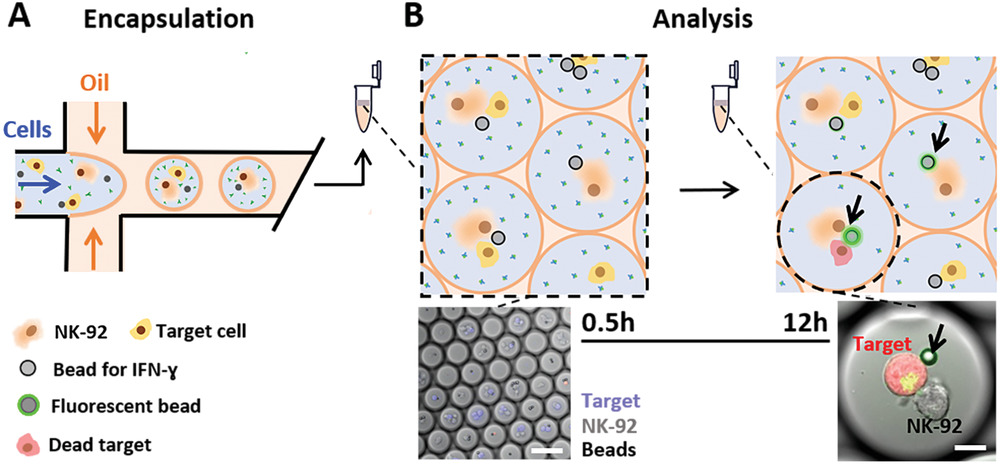
  • (A)在这个T型微流控装置中,包含NK-92细胞、靶细胞、IFN-γ感应珠和检测抗体的液滴被生成。
  • (B)将装载细胞的液滴收集到离心管中,放入恒温箱(5% CO2, 37 °C)中。在不同的时间点(0.5, 3, 6, 12 h),从自制的装载离心管收集的液滴的观察室里,通过共聚焦荧光显微镜分析IFN-γ释放和NK细胞毒性。抗体功能化的聚苯乙烯小珠用于定量分析只包含NK细胞及NK细胞和癌症细胞靶标共培养的IFN-γ分泌。DNA嵌入剂被用于评估NK细胞毒性。下面为代表性的共聚焦荧光显微图片。

Figure 2. Preparation and calibration of the IFN-γ sensing beads

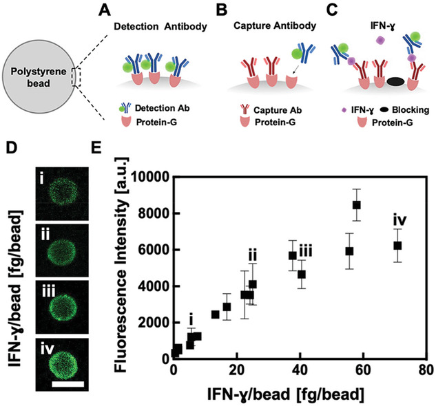
  • (A)滴定检测抗体以进行IFN-γ检测。直到饱和点为止,小珠外围的荧光强度信号根据荧光标记的检测抗体的浓度而不同,这样就可以确定传感器的最大视觉容量。
  • (B)滴定抗体以捕获IFN-γ。
  • (C)校准IFN-γ感应珠。分别对捕获抗体和检测抗体进行滴定后,将聚苯乙烯小珠(涂有4 µg mL-1捕获抗体)与确定浓度的IFN-γ(0–70 fg bead−1)以及35 µg mL-1检测抗体一起孵育3 h,液滴直径约60 µm(≈113 pl)。然后,分析20-54个小珠外围的荧光强度,并将其与各自的IFN-γ浓度相关联。
  • (D)代表性的荧光共聚焦图片,显示了小珠的中心焦点平面,揭示了小珠外围荧光强度和每个小珠被检测到的IFN-γ量的相关性。其添加的IFN-γ浓度分别为: i = 15, ii = 25, iii = 40, iv = 70 fg bead−1
  • (E)对于每个测试的IFN-γ量(如(D)给出的i,ii,iii和iv),通过共聚焦荧光显微镜检测20-54个小珠外围的荧光强度,进行分析,并将其与各自的IFN-γ量。每个数据点代表平均值±标准差。

这里的a.u.是指任意单位(Arbitrary Unit)。

Figure 3. Correlation between NK-92 cells’ IFN-γ release and their cytolytic activity

  • (A)在不同的孵育时间(0.5 h以及3、6和12 h后)下,液滴中的IFN-γ释放。描绘了每个小珠外围(左Y轴)测量的荧光强度与细胞(fg cell-1,右Y轴)释放的IFN-γ量相关。≤7.5 fg cell-1的IFN-γ释放低于传感器的检测极限,小珠外围荧光强度与背景无法区分(低于检测极限的值位于图表的灰色区域)。0.5 h的时间点表示显微镜图像采集的开始,大约与装载细胞/小珠的液滴的产生后30分钟相吻合。对于此分析的每种条件,均从相同的充满细胞的液滴群体中分析了28–50个珠子。进行了未配对的带有Welch校正的t检验(****表示p≤0.0001,***表示p≤0.001,**表示p≤0.01,ns表示p> 0.05)。在0.5和3小时时,结果没有统计学意义。
  • (B)单个液滴的代表性荧光共聚焦图像。在测定开始时(i)和孵育12小时后(ii)的NK-92细胞和IFN-γ感应珠。
  • (C)在不同时间(0.5 h以及3、6和12 h后)下,IFN-γ释放和NK-92细胞的溶细胞活性。黑点表示液滴中的NK-92细胞没有杀死靶细胞。叉代表液滴包含细胞毒性NK-92细胞,红叉代表液滴包含的细胞释放的IFN-γ量低于检测限,蓝叉的高于检测限。
  • (D)含有单个NK-92细胞、K562靶细胞和IFN-γ感应珠的液滴的代表性荧光共聚焦图片。(i)测定开始(ii)孵化5小时。死亡K562细胞被描述为红色。
  • (E)添加人重组IFN-γ(hrIFN-γ)对NK-92细胞细胞毒性的影响。NK-92细胞和K562靶细胞被悬浮在培养基分别包含0(黑条)、≈ 40(灰条)、≈ 80 fg droplet−1(白条)的hrIFN-γ中。在每种条件下,细胞被封装到50-65 µm直径的液滴,随着时间的推移(0.5 h和3、6、12 h),通过碘化丙啶(PI)染色和共聚焦荧光显微镜观察NK-92细胞的溶细胞活性。在3、6和12小时,结果没有统计学意义。

Welch校正:若假定方差齐性(各组样本具有相同的方差),则不需要Welch校正,否则则做Welch校正。

Conclusion

  通过该基于液滴的方法,作者发现在分析的早期阶段(长达6小时),释放IFN-γ的NK细胞比没有释放到可检测量的细胞因子的数量低,反映了NK细胞群在IFN-γ释放方面的异质性。在测定结束时,几乎一半被分析的活化NK-92细胞群体释放出可检测量的IFN-γ。尽管没有特定的IFN-γ释放阈值驱动NK细胞的细胞毒性,依然可以看出在12小时试验时间里,IFN-γ和NK-92细胞毒性的正向关联。

  此外也探究了外源添加人重组IFN-γ对NK-92溶细胞性的影响。暴露在高浓度的外源性IFN-γ会导致较低的NK-92细胞的细胞毒性。

Other Information

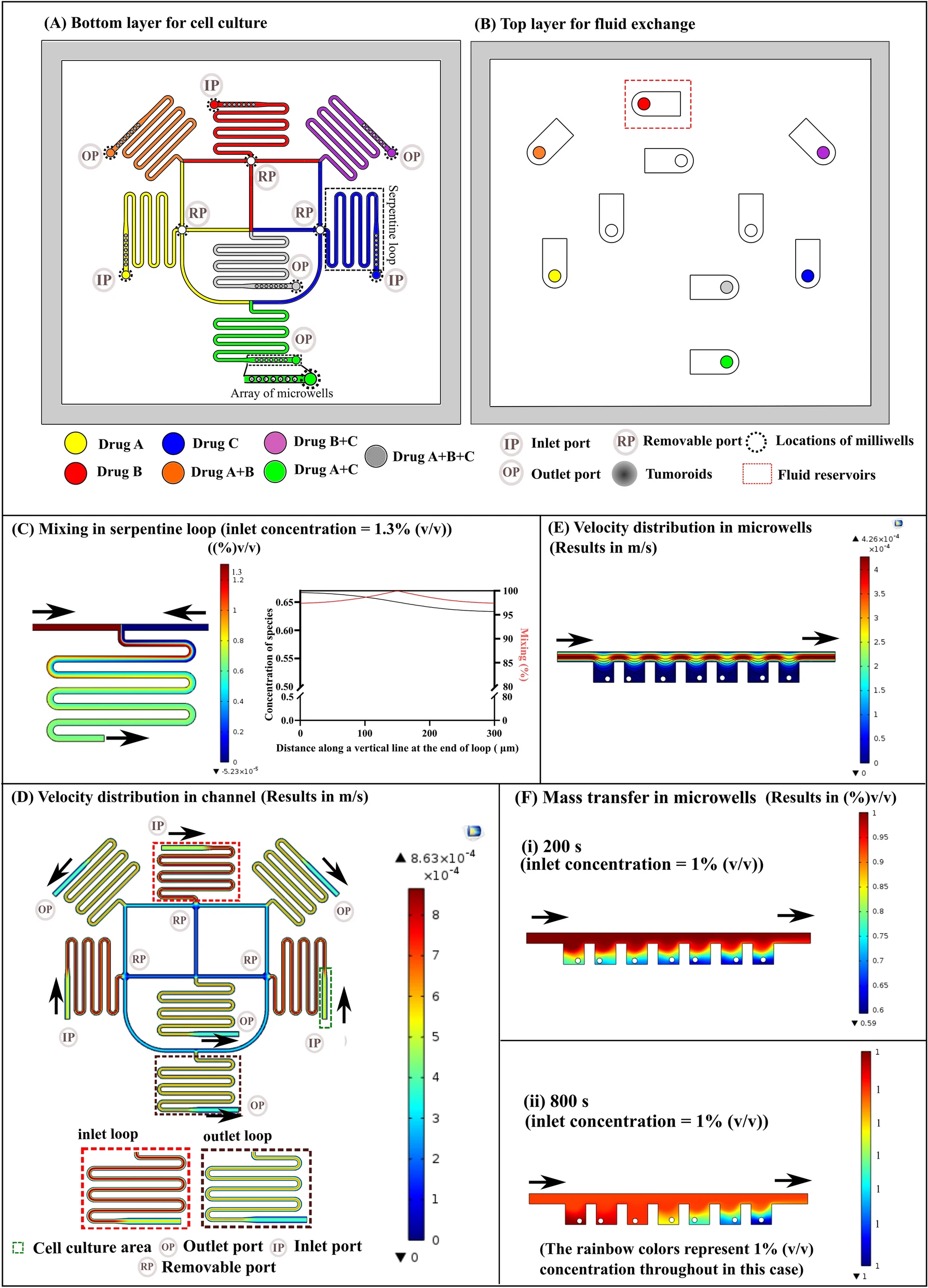
MicrofluidicChip Design

  从左至右分别为进油口、进水口和出口。

Reference

Antona S, Abele T, Jahnke K, et al. Droplet-Based Combinatorial Assay for Cell Cytotoxicity and Cytokine Release Evaluation[J]. Advanced Functional Materials, , n/a(n/a): 2003479.

评论

3D cell culture 3D cell culturing 3D cell microarrays 3D culture 3D culture model 3D printing 3D spheroid 3D tumor culture 3D tumors 3D vascular mapping ACT ADV AUTODESK Abdominal wall defects Acoustofluidics Adipocyte Adipogenesis Adoptive cell therapy AirPods Alginate Anticancer Anticancer agents Anticancer drugs Apple Apriori Association Analysis August AutoCAD Autodock Vina Bio-inspired systems Biochannels Bioengineering Bioinspired Biological physics Biomarkers Biomaterial Biomaterials Biomimetic materials Biomimetics Bioprinting Blood purification Blood-brain barrier Bone regeneration Breast cancer Breast cancer cells Breast neoplasms CM1860 CRISPR/Cas9 system CSS CTC isolation CTCs Cancer Cancer angiogenesis Cancer cell invasion Cancer immunity Cancer immunotherapy Cancer metabolism Cancer metastasis Cancer models Cancer screening Cancer stem cells Cell adhesion Cell arrays Cell assembly Cell clusters Cell culture Cell culture techniques Cell mechanical stimulation Cell morphology Cell trapping Cell-bead pairing Cell-cell interaction Cell-laden gelatin methacrylate Cellular uptake Cell−cell interaction Cervical cancer Cheminformatics Chemotherapy Chimeric antigen receptor-T cells Chip interface Circulating tumor cells Clinical diagnostics Cmder Co-culture Coculture Colon Colorectal cancer Combinatorial drug screening Combinatorial drug testing Compartmentalized devices Confined migration Continuous flow Convolutional neural network Cooking Crawler Cryostat Curved geometry Cytokine detection Cytometry Cytotoxicity Cytotoxicity assay DESeq DNA tensioners Data Mining Deep learning Deformability Delaunay triangulation Detective story Diabetic wound healing Diagnostics Dielectrophoresis Differentiation Digital microfluidics Direct reprogramming Discrimination of heterogenic CTCs Django Double emulsion microfluidics Droplet Droplet microfluidics Droplets generation Droplet‐based microfluidics Drug combination Drug efficacy evaluation Drug evaluation Drug metabolism Drug resistance Drug resistance screening Drug screening Drug testing Dual isolation and profiling Dynamic culture Earphone Efficiency Efficiency of encapsulation Elastomers Embedded 3D bioprinting Encapsulation Endothelial cell Endothelial cells English Environmental hazard assessment Epithelial–mesenchymal transition Euclidean distance Exosome biogenesis Exosomes Experiment Extracellular vesicles FC40 FP-growth Fabrication Fast prototyping Fibroblasts Fibrous strands Fiddler Flask Flow rates Fluorescence‐activated cell sorting Functional drug testing GEO Galgame Game Gene Expression Profiling Gene delivery Gene expression profiling Gene targetic Genetic association Gene‐editing Gigabyte Glypican-1 GoldenDict Google Translate Gradient generator Growth factor G‐CSF HBEXO-Chip HTML Hanging drop Head and neck cancer Hectorite nanoclay Hepatic models Hepatocytes Heterotypic tumor HiPSCs High throughput analyses High-throughput High-throughput drug screening High-throughput screening assays High‐throughput methods Histopathology Human neural stem cells Human skin equivalent Hydrogel Hydrogel hypoxia Hydrogels ImageJ Immune checkpoint blockade Immune-cell infiltration Immunoassay Immunological surveillance Immunotherapy In vitro tests In vivo mimicking Induced hepatocytes Innervation Insulin resistance Insulin signaling Interferon‐gamma Intestinal stem cells Intracellular delivery Intratumoral heterogeneity JRPG Jaccard coefficient JavaScript July June KNN Kidney-on-a-chip Lab-on-a-chip Laptop Large scale Lattice resoning Leica Leukapheresis Link Lipid metabolism Liquid biopsy Literature Liver Liver microenvironment Liver spheroid Luminal mechanics Lung cells MOE Machine Learning Machine learning Macro Macromolecule delivery Macroporous microgel scaffolds Magnetic field Magnetic sorting Malignant potential Mammary tumor organoids Manhattan distance Manual Materials science May Mechanical forces Melanoma Mesenchymal stem cells Mesoporous silica particles (MSNs) Metastasis Microassembly Microcapsule Microcontact printing Microdroplets Microenvironment Microfluidic array Microfluidic chips Microfluidic device Microfluidic droplet Microfluidic organ-on-a chip Microfluidic organ-on-a-chip Microfluidic patterning Microfluidic screening Microfluidic tumor models Microfluidic-blow-spinning Microfluidics Microneedles Micropatterning Microtexture Microvascular Microvascular networks Microvasculatures Microwells Mini-guts Mirco-droplets Molecular docking Molecular imprinting Monolith Monthly Multi-Size 3D tumors Multi-organoid-on-chip Multicellular spheroids Multicellular systems Multicellular tumor aggregates Multi‐step cascade reactions Myeloid-derived suppressor cells NK cell NanoZoomer Nanomaterials Nanoparticle delivery Nanoparticle drug delivery Nanoparticles Nanowell Natural killer cells Neural progenitor cell Neuroblastoma Neuronal cell Neurons Nintendo Nissl body Node.js On-Chip orthogonal Analysis OpenBabel Organ-on-a-chip Organ-on-a-chip devices Organically modified ceramics Organoids Organ‐on‐a‐chip Osteochondral interface Oxygen control Oxygen gradients Oxygen microenvironments PDA-modified lung scaffolds PDMS PTX‐loaded liposomes Pain relief Pancreatic cancer Pancreatic ductal adenocarcinoma Pancreatic islet Pathology Patient-derived organoid Patient-derived tumor model Patterning Pearl powder Pearson coefficient Penetralium Perfusable Personalized medicine Photocytotoxicity Photodynamic therapy (PDT) Physiological geometry Pluronic F127 Pneumatic valve Poetry Polymer giant unilamellar vesicles Polystyrene PowerShell Precision medicine Preclinical models Premetastatic niche Primary cell transfection Printing Protein patterning Protein secretion Pubmed PyMOL Pybel Pytesseract Python Quasi-static hydrodynamic capture R RDKit RNAi nanomedicine RPG Reactive oxygen species Reagents preparation Resistance Review Rod-shaped microgels STRING Selective isolation Self-assembly Self-healing hydrogel September Signal transduction Silk-collagen biomaterial composite Similarity Single cell Single cells Single molecule Single-cell Single-cell RNA sequencing Single‐cell analysis Single‐cell printing Size exclusion Skin regeneration Soft lithography Softstar Spheroids Spheroids-on-chips Staining StarBase Stem cells Sub-Poisson distribution Supramolecular chemistry Surface chemistry Surface modification Switch T cell function TCGA Tanimoto coefficient The Millennium Destiny The Wind Road Thin gel Tissue engineering Transcriptome Transfection Transient receptor potential channel modulators Tropism Tubulogenesis Tumor environmental Tumor exosomes Tumor growth and invasion Tumor immunotherapy Tumor metastasis Tumor microenvironment Tumor response Tumor sizes Tumor spheroid Tumor-on-a-chip Tumorsphere Tumors‐on‐a‐chip Type 2 diabetes mellitus Ultrasensitive detection Unboxing Underlying mechanism Vascularization Vascularized model Vasculature Visual novel Wettability Windows Terminal Word Writing Wuxia Xenoblade Chronicles Xin dynasty XuanYuan Sword Youdao cnpm fsevents miR-125b-5p miR-214-3p miRNA signature miRanda npm
Your browser is out-of-date!

Update your browser to view this website correctly. Update my browser now

×